

Cynthia A. Araradian, MD
Resident Physician
OHSU
Portland, OR, United States
All of the relevant financial relationships listed below have been mitigated.
This individual has no financial relationships with ineligible companies.

Sandy H. Fang, MD
Associate Professor
Oregon Health and Science University
Portland, Oregon, United States
All of the relevant financial relationships listed below have been mitigated.
This individual has no financial relationships with ineligible companies.
Rebekka Duhen, Ph.D.
Senior Scientist
Cancer Early Detection Advanced Research Center (CEDAR), OHSU Knight Cancer Institute
Portland, Oregon, United States
Disclosure information not submitted.
Mariah Erlick, MD
Surgical Resident
OHSU
Portland, Oregon, United States
Disclosure information not submitted.
Cynthia A. Araradian, MD
Resident Physician
OHSU
Portland, OR, United States
All of the relevant financial relationships listed below have been mitigated.
This individual has no financial relationships with ineligible companies.
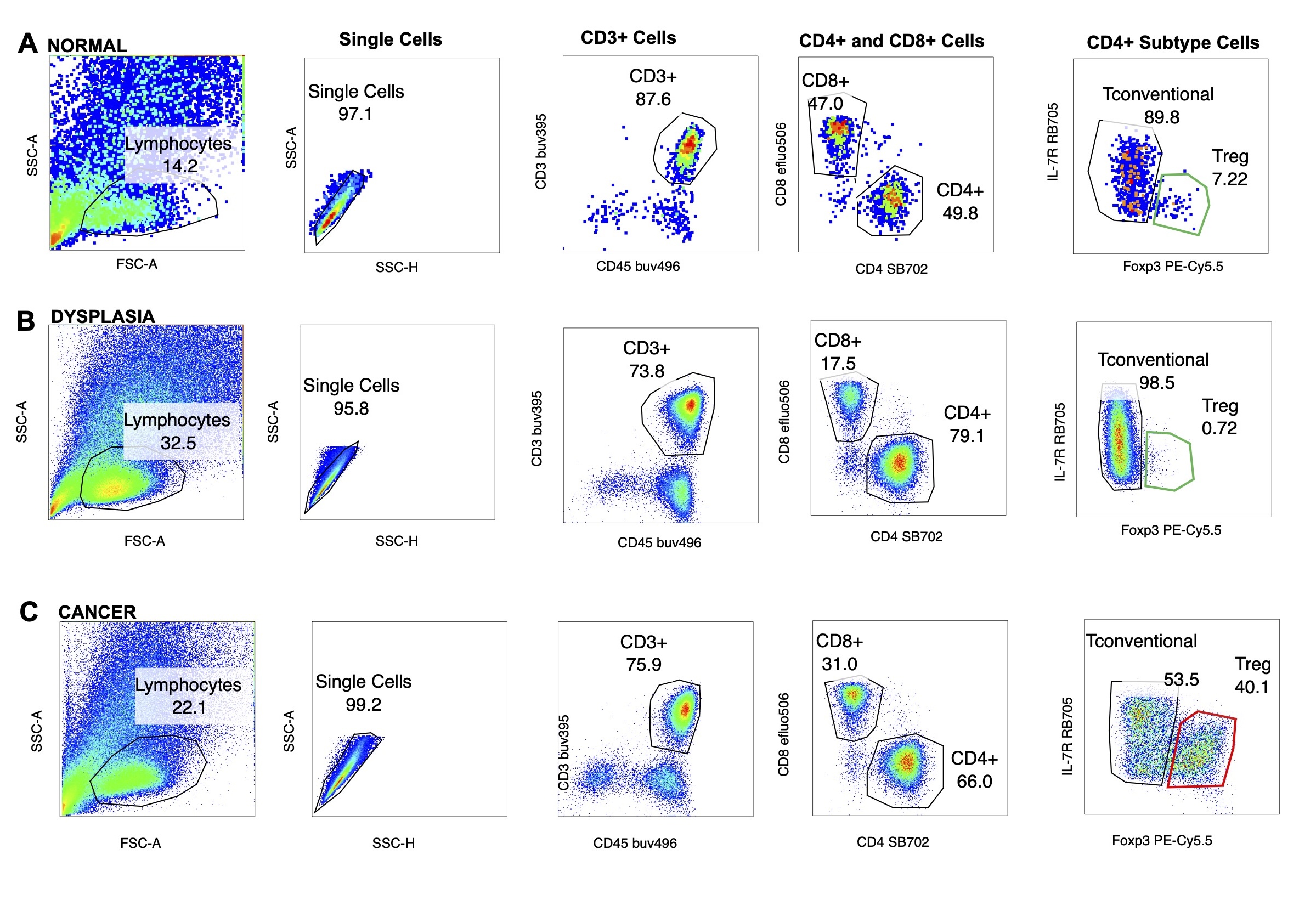 (A) Flow cytometric analysis for CD4 and CD8 T cells including the gating strategy for the normal tissue specimen. The normal specimen has overall lower lymphocyte infiltration. There are very few regulatory T cells (Tregs) outlined in green. (B) Flow cytometric analysis and gating strategy for the tissue with dysplasia. There are more lymphocytes present within the tissue sample but very few Tregs are noted within the CD4 T cell populations. (C) Flow cytometric analysis and gating strategy for anal squamous cell carcinoma. There are significantly more Treg cells present within the CD4 subset. The Treg cells are identified by the FOXP3 stain outlined above in red.
 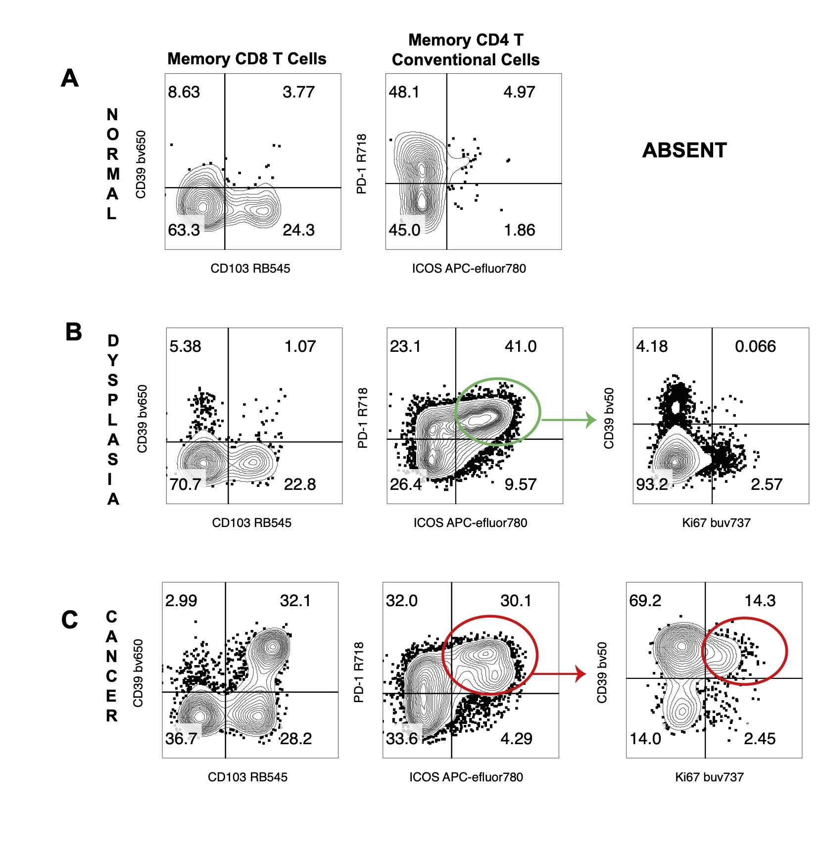 (A) Flow cytometric analysis of the normal specimen contains very few CD103+ CD39+ [double positive (DP)] memory CD8+ T cells and very few DP ICOS+ PD-1+ CD4 T cells. Given the lack of double positive cells further analysis was not performed for activation markers. (B) Flow cytometric analysis of the tissue with dysplasia contains very few DP CD103+ CD39+ memory CD8+ T cells. There are a large proportion of DP ICOS+ PD-1+ CD4 T cells within this population, outlined in green, but once the DP cells are gated they do not appear to express the activation marker Ki-67. (C) Flow cytometric analysis of the cancer specimen demonstrates a higher presence of both DP CD103+ CD39+ CD8 and DP ICOS+ PD-1+ CD4 T cells. Gating the DP ICOS+ PD-1+ CD4 T cells demonstrates higher expression of activation markers, Ki-67 and CD39, outlined in red.